
During early embryonic development, cells have to make two things happen at the same time: they need to divide rapidly to increase the number of cells, while also changing their identity and organisation to build an embryo.
One of the central signalling pathways controlling this transition is the FGF–MEK–ERK pathway. ERK signalling helps embryonic cells leave the naïve state of pluripotency and progress towards more differentiated developmental states. But while studying this process, we wondered whether the cell itself—not only signals coming from outside the cell—could also influence ERK activity.
To address this question, we developed a large-scale experimental approach using embryonic stem cells as a model for the early epiblast and searched for previously unknown regulators of embryonic development.
Looking inside the cell for regulators of ERK
Around the time of implantation, the mouse embryo undergoes a remarkable transformation. The epiblast, which initially consists of a small group of pluripotent cells, rapidly expands and reorganises into an epithelial structure. At the same time, the cells transition from naïve pluripotency towards a more developmentally advanced state.
We recreated aspects of this transition using embryonic stem cells and developed an automated phenotypic screening system. This allowed us to systematically interfere with cellular pathways and ask which factors influence the transition out of naïve pluripotency.
Among the strongest hits was Aurora kinase A (AURKA).
AURKA is best known as a kinase involved in cell division, particularly in the organisation of centrosomes and the mitotic spindle. Its identification was therefore unexpected: why would a protein classically associated with mitosis influence ERK signalling and developmental progression?
AURKA connects cell division to developmental signalling
We found that AURKA contributes to ERK activation in cells that are not undergoing division. When we inhibited AURKA, ERK activity decreased and the transcriptional programme normally associated with exiting naïve pluripotency was delayed.
In other words, AURKA acts as a cell-intrinsic regulator of ERK signalling, working alongside the better-known FGF–MEK pathway.
This finding suggests that the developmental state of a cell is not controlled simply by signals that arrive from its environment. Instead, the cell’s own internal machinery can also influence whether it is ready to progress developmentally.
This is particularly interesting in the early embryo, where cells are simultaneously receiving developmental signals and undergoing extremely rapid cycles of cell division.
But what happens to ERK when a cell divides?
This observation led us to another question.
When a cell enters mitosis, transcription largely stops. Therefore, the classical role of ERK—controlling gene expression—cannot operate in the same way.
So where does ERK go during cell division?
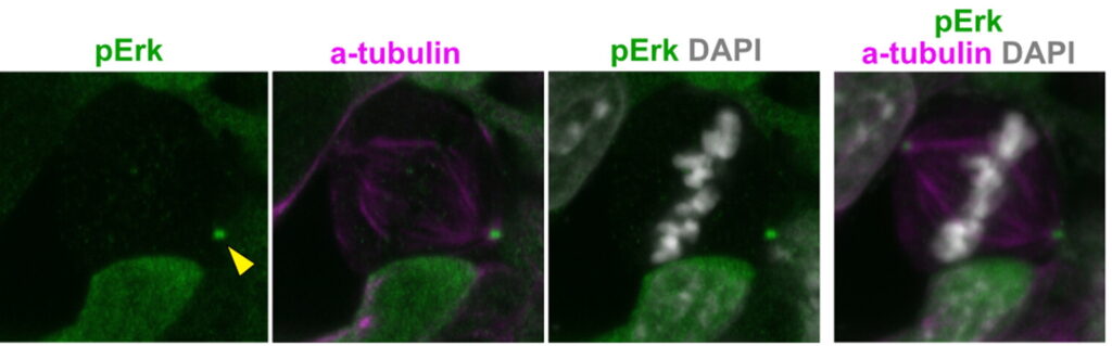
Using high-resolution imaging, we found that ERK changes its localisation when cells enter mitosis. Following nuclear envelope breakdown, phosphorylated ERK accumulated at the centrosomes and spindle poles, where it overlapped with components of the mitotic machinery.
This was particularly striking because it suggested that ERK may have a function during mitosis that is fundamentally different from its classical role as a transcriptional regulator.
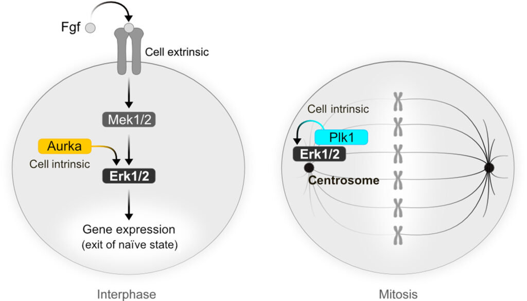
Erk is involved in two modes of activity in relation to the cell cycle progression. In interphase cells, Erk drives a transcriptional response that is promoted by cell-extrinsic (Fgf/Mek) and intrinsic (Aurka) signaling. During mitosis, as transcription stops, Erk positions at the centrosomes, where its activity depends on Plk1.
A second mitotic kinase enters the picture
We next investigated how ERK becomes activated at the centrosomes during mitosis.
Our experiments pointed to Polo-like kinase 1 (PLK1), another major regulator of cell division. Inhibition of PLK1 reduced ERK phosphorylation at the centrosomes, indicating that PLK1 controls ERK activation at this location.
Together, our findings reveal an unexpected relationship between two types of signalling:
During interphase:
FGF → MEK → ERK → transcription → developmental progression
with AURKA providing an additional cell-intrinsic input into ERK activation.
During mitosis:
PLK1 → ERK phosphorylation → centrosomal localisation
Here, transcription is switched off and ERK becomes associated with the machinery that controls cell division.
ERK has two different lives
Our work therefore suggests that ERK is not simply an on/off developmental signalling molecule.
Instead, its function changes with the cell-cycle state of the cell.
During interphase, ERK can enter the nucleus and regulate gene expression, helping cells leave naïve pluripotency and progress towards the next stage of development.
When the cell enters mitosis, transcription shuts down and ERK is redirected to the centrosomes, where its activation depends on PLK1.
We do not yet know exactly what ERK does at the centrosomes during mitosis. This remains an intriguing open question. It is possible that ERK contributes directly to aspects of the cell-division machinery, but further work will be needed to determine its precise function.
Why does this matter for embryonic development?
The early embryo faces a remarkable coordination problem. Its cells must divide rapidly, maintain or change their developmental identity at the appropriate time, and reorganise themselves into increasingly complex structures.
Our findings suggest that these processes may be more tightly connected than previously appreciated.
Rather than treating cell division and developmental signalling as separate processes, our work reveals a molecular connection between them. The same ERK pathway that helps determine the developmental state of a cell is also recruited to the machinery that drives cell division.
This provides a new perspective on how embryonic cells coordinate proliferation, signalling and developmental progression.
More broadly, it highlights an important principle: developmental decisions are shaped not only by signals coming from the outside, but also by the internal state of the cell—including the machinery that controls when and how a cell divides.
In short, we found that the mitotic kinases AURKA and PLK1 provide cell-intrinsic inputs into ERK signalling, revealing two distinct modes of ERK activity: one that controls developmental gene expression during interphase and another associated with the cell-division machinery during mitosis.
Read the original article:
Developmental regulation of Erk signaling by mitotic kinases
Chen F, Bruder JM, Govindasamy N, Wu J, Chen R, Prit D, Drexler HCA, Leidel SA, Schöler HR, Bedzhov I
Science Advances 2026
https://doi.org/10.1126/sciadv.adq7469