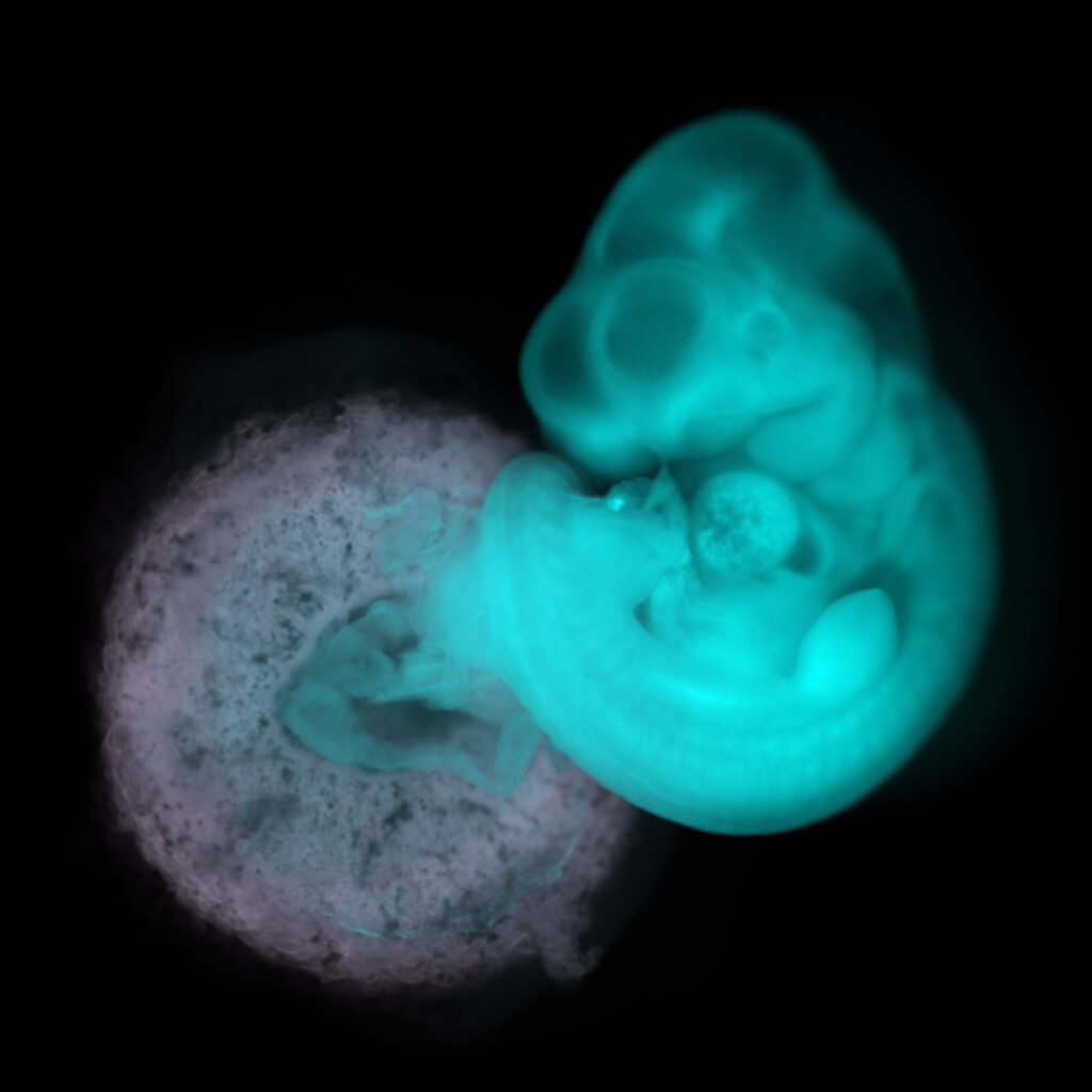
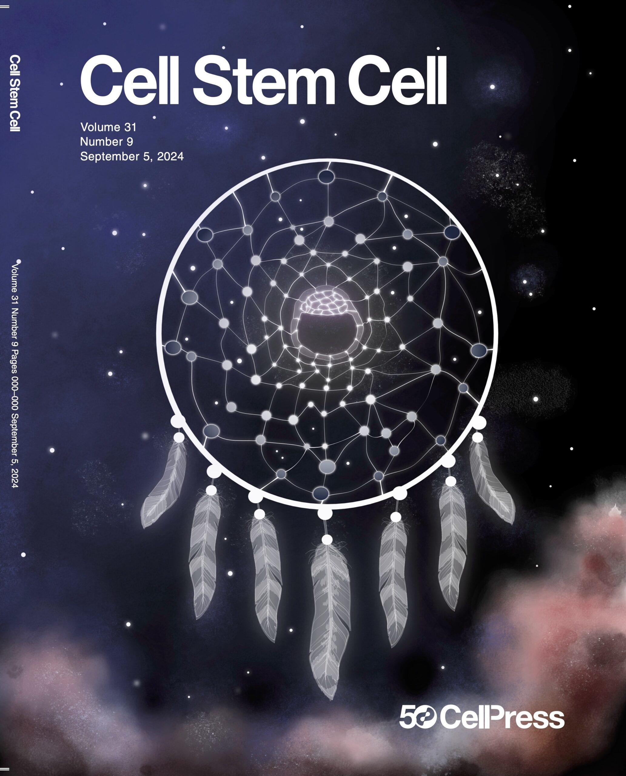
COVERS


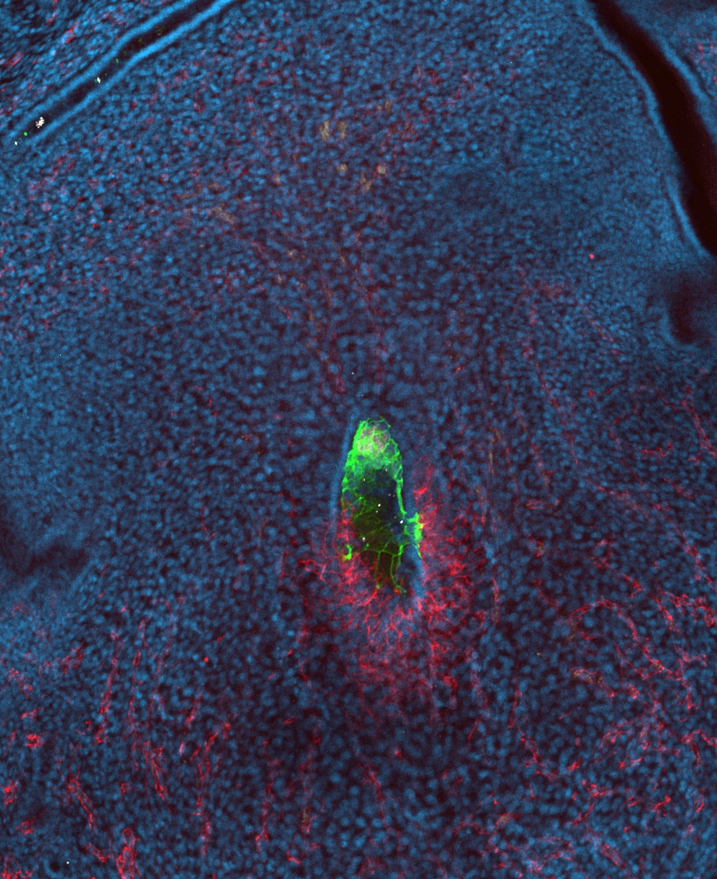
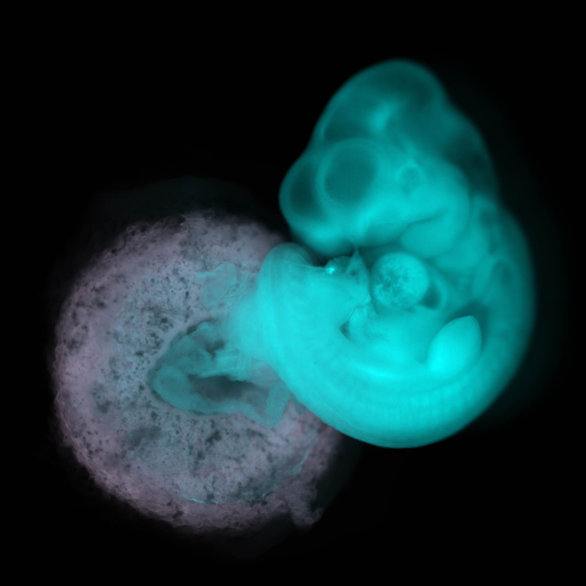
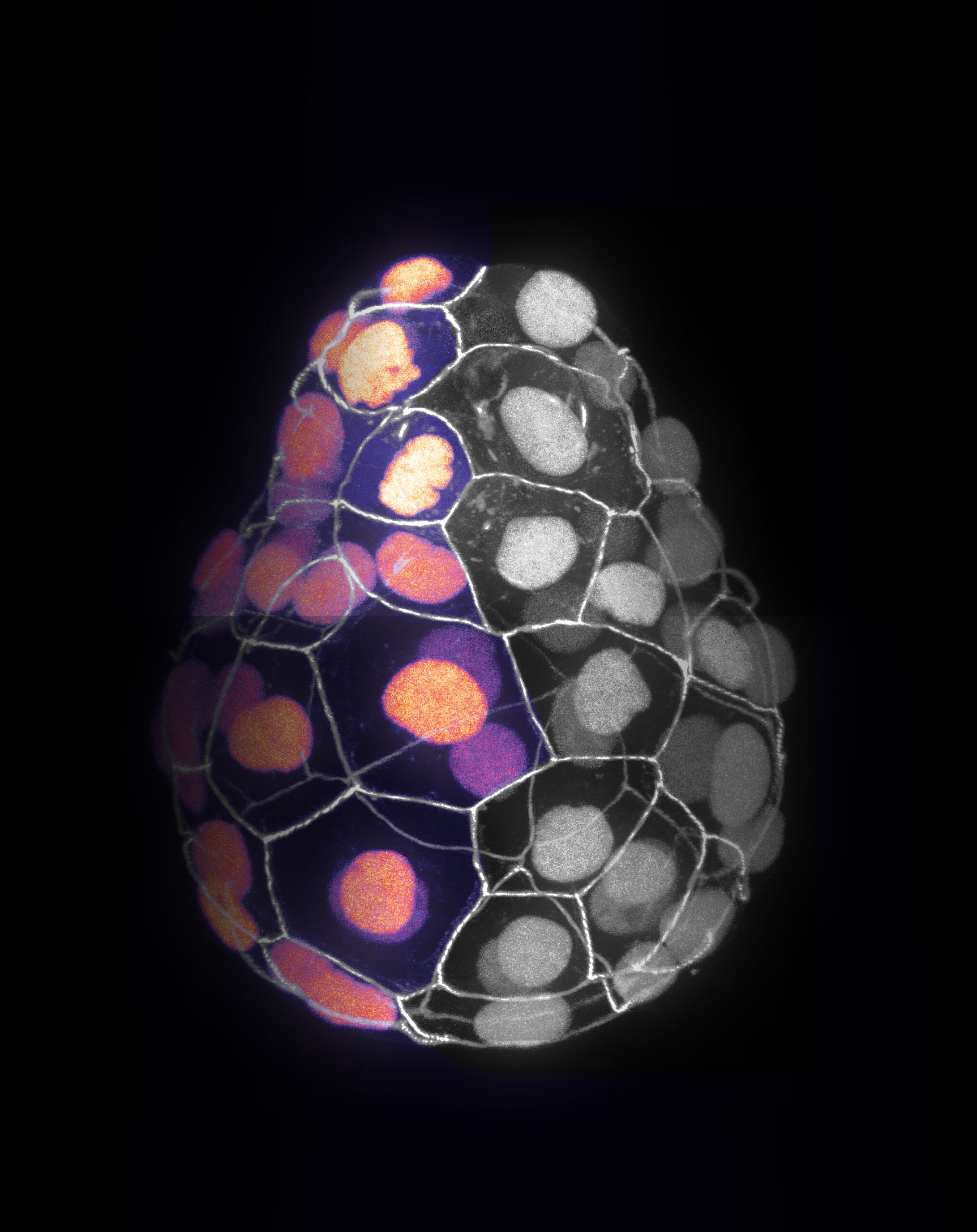
MICROSCOPY IMAGES AND CANDIDATE COVERS

MOVIES
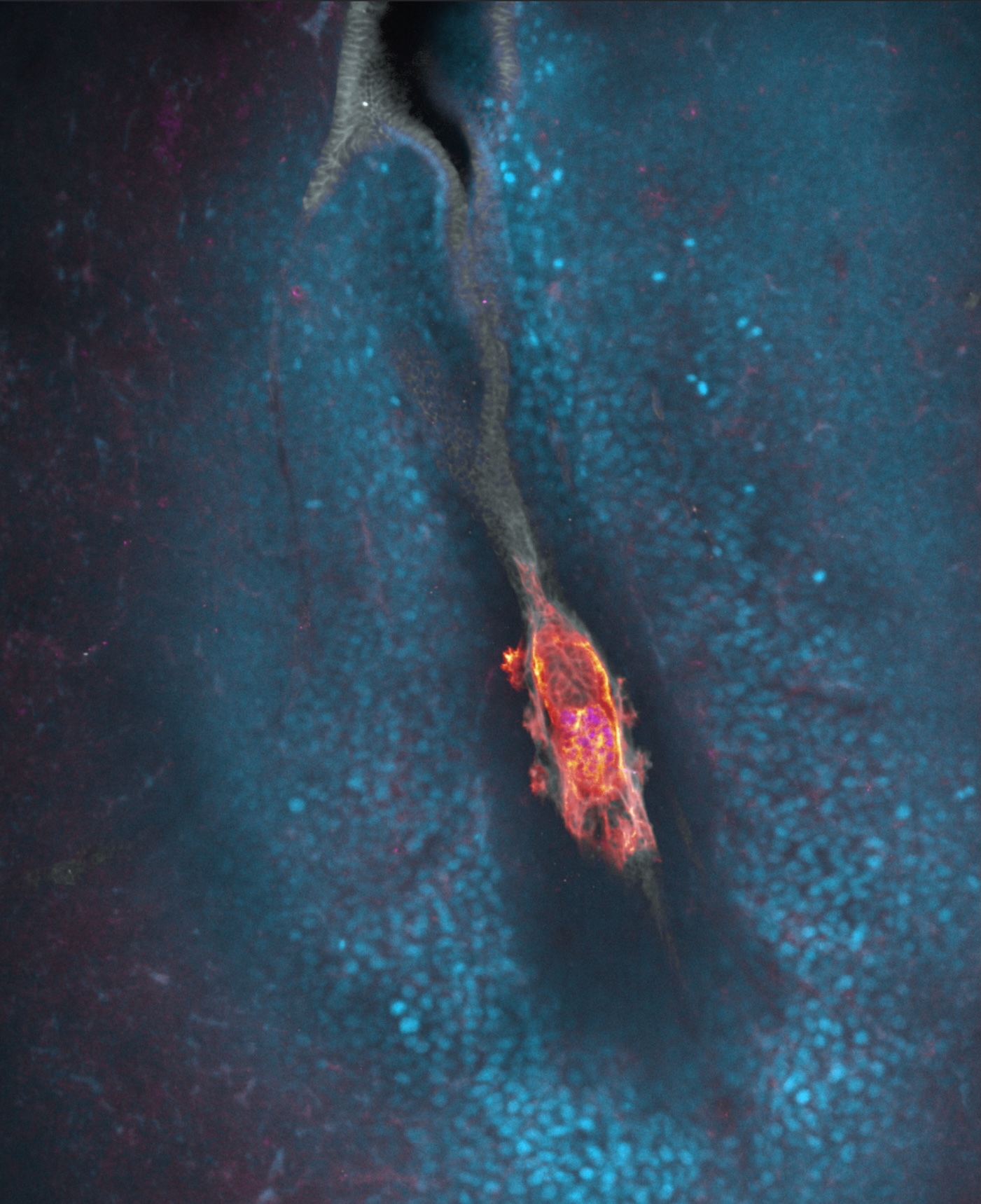
3D biomimetic environment enabling embryo “implantation” in vitro
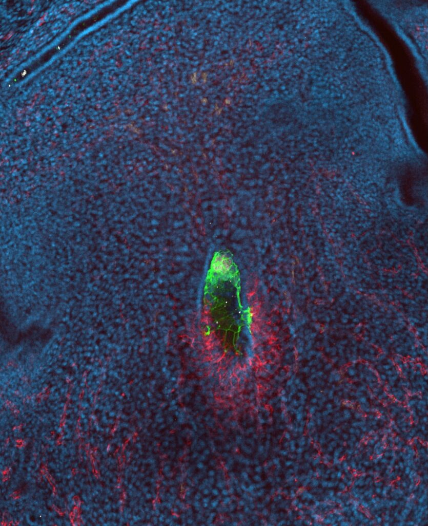
Mouse embryo and endothelial cells interactions in vitro, in a microfluidic chip (Govindasamy et al, Developmental Cell 2021 https://doi.org/10.1016/j.devcel.2021.10.014)
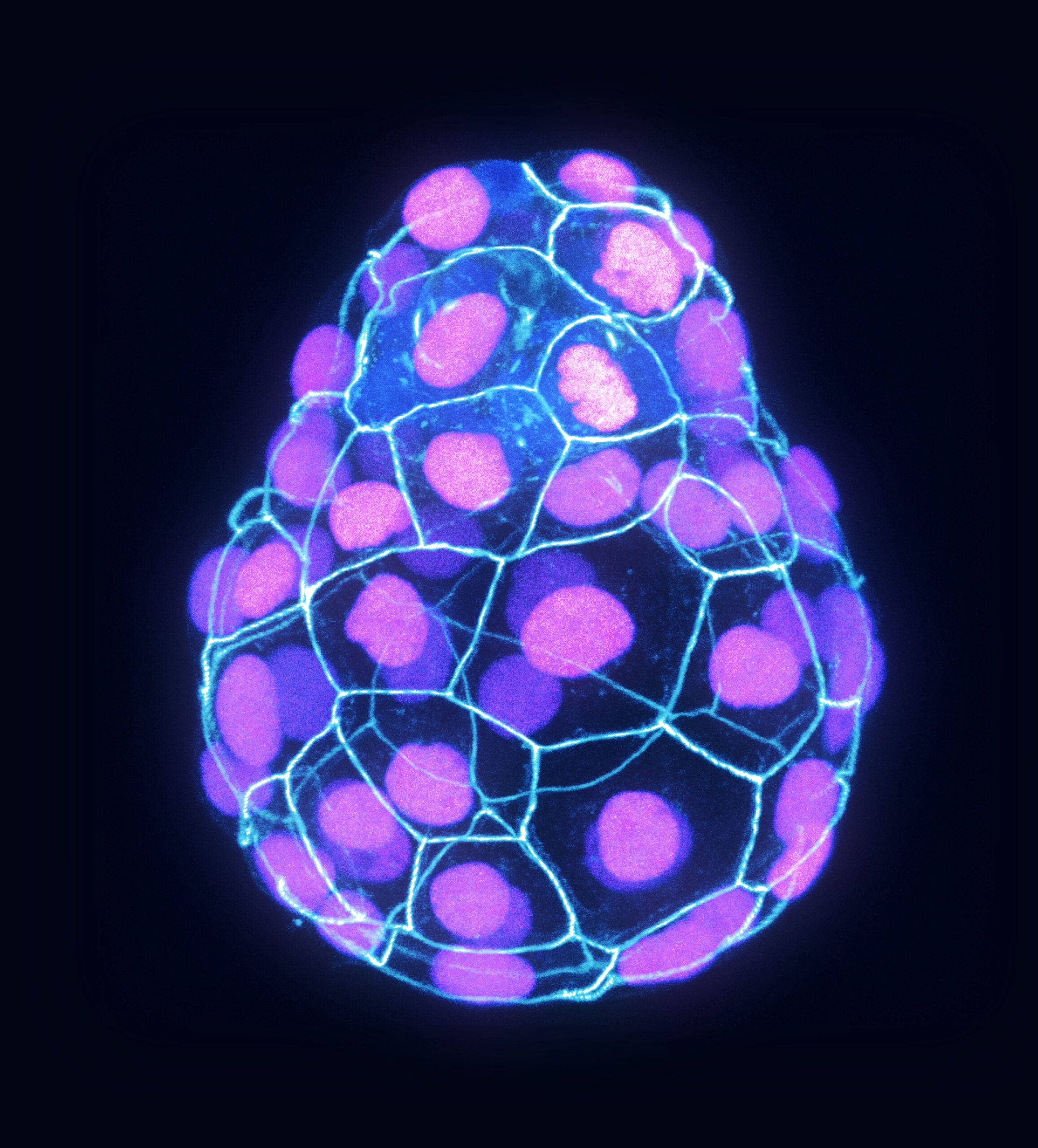
In vitro model of the proamniotic cavity formation
Serial block face scanning electron microscopy of mouse pluripotent cells grown in 3D culture conditions. The central lumen and the intermembranous spaces near the lumen are 3D rendered.
Kim et al., Science Advances 2022 https://doi.org/10.1126/sciadv.abe1640
OVERVIEW AND MORE

“In the beginning there was nothing, which exploded.”
You must be logged in to post a comment.