
During early embryonic development, the cells that will eventually form the placenta undergo a remarkable transformation. A small population of cells at the embryonic pole of the blastocyst gives rise to the trophoblast stem-cell compartment, which subsequently expands and develops into the extra-embryonic tissues that support the growing embryo.
But how does this relatively simple layer of cells reorganise itself into the complex architecture of the developing placenta?
We wanted to understand how this transformation takes place during the transition from the blastocyst to the early post-implantation embryo.
A surprising change in tissue architecture
At the blastocyst stage, the polar trophectoderm has a typical epithelial organisation, with its apical and basal surfaces oriented towards defined sides of the tissue.
As development progresses, however, the trophoblast undergoes a dramatic reorganisation.
The prevailing model suggested that this transformation was largely driven by tissue folding. Our observations led us to a different explanation.
We found that the trophoblast stem-cell compartment is reorganised through an inversion of epithelial polarity.
Rather than simply folding an existing epithelial layer, the tissue changes the orientation of its polarity axis. This large-scale rearrangement allows the trophoblast stem-cell compartment to adopt a new architecture as the embryo transitions from the blastocyst to the egg-cylinder stage.
Turning the polarity axis around
Epithelial cells are highly organised. Their apical and basal surfaces have different molecular compositions and functions, and this polarity determines how cells interact with their neighbours and their surrounding environment.
We found that this polarity is dramatically remodelled during trophoblast development.
The extracellular matrix and basement membrane provide important positional information to the cells. As the tissue reorganises, the polarity cues transmitted through the basement membrane contribute to the new orientation of the trophoblast cells.
In particular, we found that β1, β3 and β5 integrins work together to transmit these polarising signals.
This means that the extracellular environment does not simply provide structural support. It actively contributes to determining how the trophoblast tissue is organised.
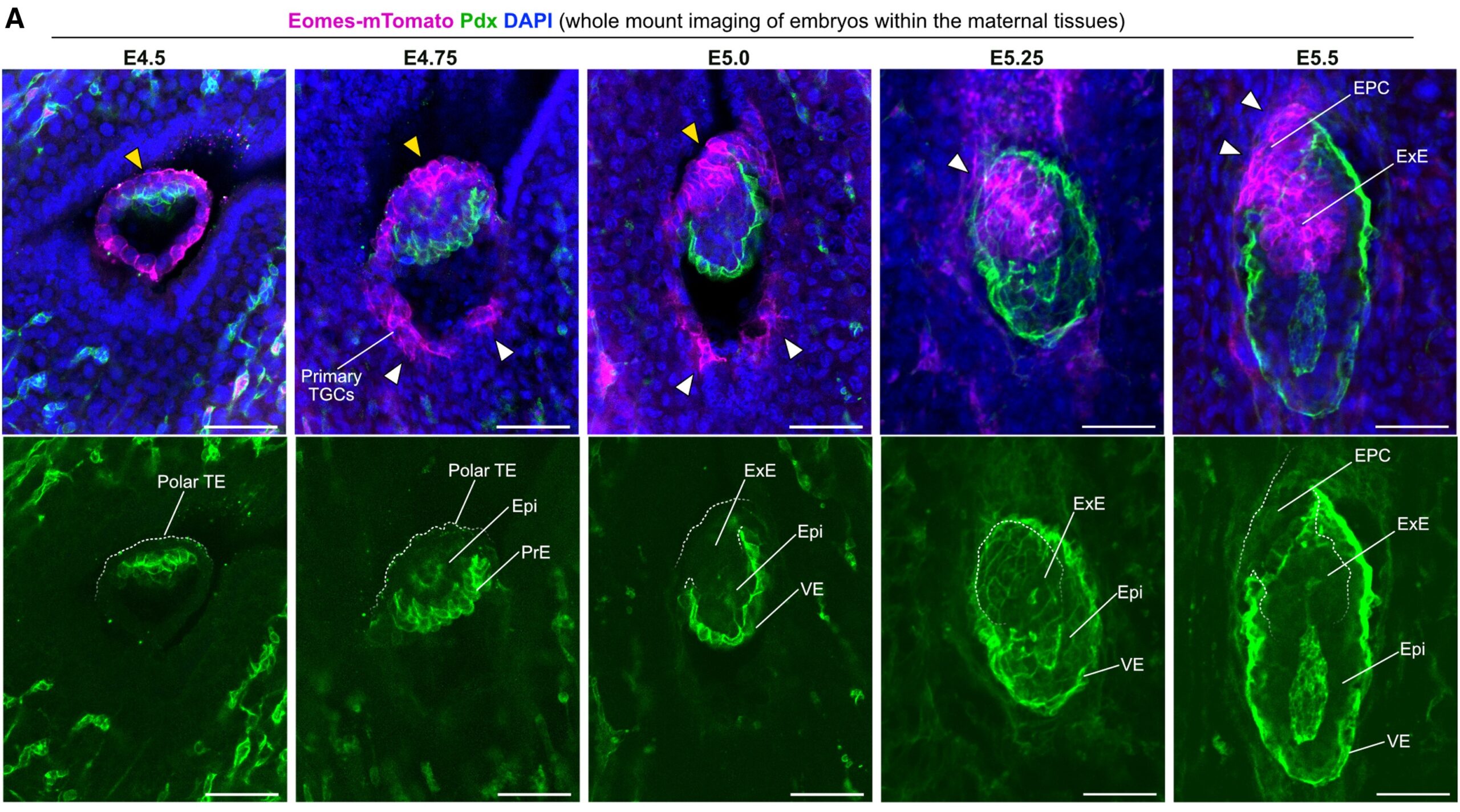
Representative images of whole-mount staining of uterine tissues containing E4.5, E4.74, E5.0, E5.25, and E5.5 embryos. White arrowheads indicate the formation of the embryo-maternal interface, yellow arrowheads indicate the E4.5, E4.75 polar TE, and posterior trophoblast cells of the E5.0 ExE.
Orthogonal transition through z-stack of tissue-cleared decidua containing an E5.0 embryo.
Orthogonal transition through z-stack of tissue-cleared decidua containing an E5.5 embryo.
Recreating the process outside the embryo
To understand whether polarity inversion was an intrinsic property of trophoblast cells, we turned to trophoblast stem cells in vitro.
Remarkably, we were able to reproduce key aspects of the polarity reorganisation outside the embryo.
This demonstrated that trophoblast cells have the capacity to respond to their surrounding environment and reorganise their polarity at the tissue scale.
By combining embryonic analysis, imaging, ultrastructural approaches and engineered extracellular environments, we could therefore connect the behaviour of individual cells with the large-scale architecture of the developing tissue.
Why does polarity matter?
The trophoblast is not simply a passive layer surrounding the embryo. It is the source of the extra-embryonic tissues that will form much of the placenta and is essential for establishing the interface between the embryo and the mother.
Its architecture therefore needs to be precisely coordinated with implantation and subsequent embryonic development.
Our findings suggest that changing the orientation of cell polarity provides a mechanism for reorganising the trophoblast stem-cell compartment without requiring extensive tissue folding.
More broadly, this work highlights how developmental tissues can change their architecture by modifying the relationship between cells and their surrounding extracellular matrix.
From cells to tissues
One of the broader lessons from this work is that tissue morphogenesis cannot always be understood simply by looking at how individual cells move.
Cells can also reorganise a tissue by changing their polarity, their interactions with the extracellular matrix and the orientation of their epithelial organisation.
We found that polarity inversion provides a mechanism for the large-scale reorganisation of the trophoblast during one of the most important transitions in early embryonic development.
In short, we discovered that the trophoblast stem-cell compartment is reorganised not simply by folding, but by turning its epithelial polarity axis around—a process in which integrin-mediated interactions with the extracellular matrix provide important polarising cues.
This provides a new framework for understanding how the placenta-forming tissues are shaped during the earliest stages of implantation.
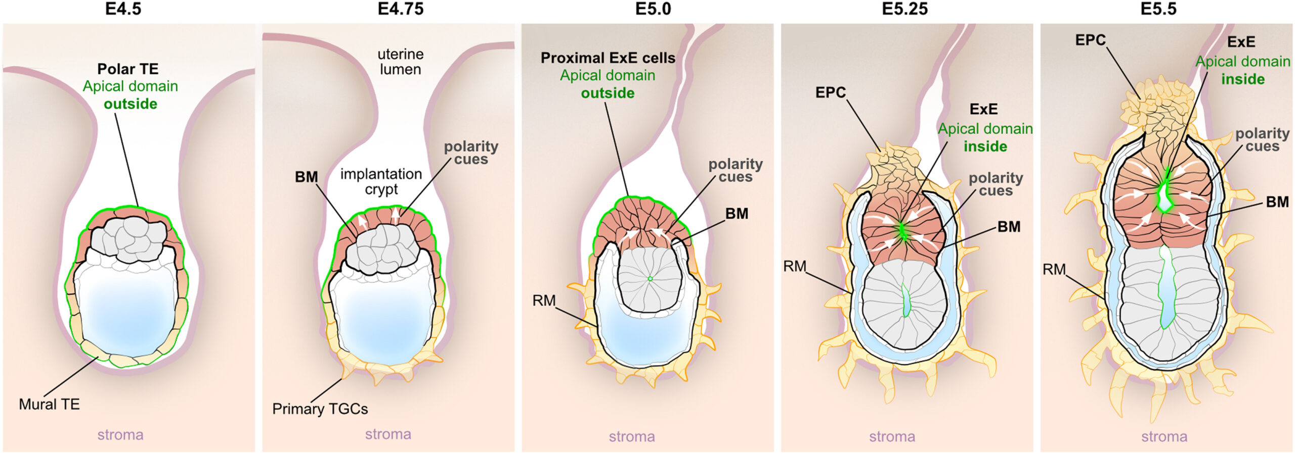
At the blastocyst stage, the trophoblast is organized as an epithelial monolayer. Upon attachment to the uterine wall (E4.5–E4.75), the distal portion of the TE (mural TE) differentiates into TGCs, which lose their epithelial phenotype. At E5.0 only the most proximal cells of the ExE still face the outside environment, exhibiting their apical membrane domains toward the open space of the implantation crypt. This is where the second site of direct embryo-maternal interactions is formed between E5.0 and E5.5, as the proximal cells of the ExE give rise to the EPC. Thus, to establish the early embryo-maternal interface, the trophoblast cells undergo differentiation, which is associated with loss of epithelial morphology.
Read the research article:
Polarity inversion reorganizes the stem cell compartment of the trophoblast lineage
Ozguldez HO, Govindasamy N, Fan R, Long H, Mildner K, Zeuschner D, Trappmann B, Ranga A, Bedzhov I.
Cell Reports 2023
https://doi.org/10.1016/j.celrep.2023.112313

You must be logged in to post a comment.