
The placenta is essential for embryonic development, yet studying how individual genes control its formation has been surprisingly difficult.
The reason is simple: the placenta and the embryo come from different cell lineages, but most of the genetic tools we use in mice are designed to manipulate the embryo as a whole.
We wanted to find a way around this problem.
In this study, we developed a simple method that allows us to genetically modify the trophoblast lineage — the cells that will form the fetal part of the placenta — directly in the early mouse embryo. Our approach uses a cell-permeable version of the Cre recombinase enzyme called Tat-Cre, together with the natural properties of the blastocyst to selectively edit genes in the trophectoderm without altering the embryonic lineage.
Why is the placenta so difficult to study?
The placenta begins with a small population of cells at the outside of the blastocyst called the trophectoderm.
These cells have a very different fate from the inner cell mass. While the inner cell mass generates the embryo proper, the trophectoderm gives rise to the trophoblast lineage and ultimately forms much of the fetal part of the placenta.
This separation is established remarkably early — but it creates a problem for genetic experiments.
To understand what a gene does in the placenta, we would ideally like to remove it from the trophoblast while leaving the embryo itself untouched.
The standard solution is the Cre/loxP system. A gene is flanked by loxP sites, and Cre recombinase removes the DNA between them. If Cre is expressed specifically in the trophoblast, we can therefore generate a trophoblast-specific knockout.
The problem was that there was no convenient Cre mouse line that provided the required specificity at the earliest trophoblast stage. Some genes that are specific to the trophectoderm in the blastocyst later become active in other tissues, raising the possibility of unwanted genetic recombination outside the placenta.
So we asked a different question:
What if we don’t need a Cre mouse at all?
Turning the blastocyst’s barrier into an advantage
The blastocyst has an interesting physical property.
Its outer layer of trophectoderm forms a barrier around the inner cell mass. This barrier is not equally permeable to everything.
We realised that we could exploit this natural property to deliver Cre directly to the cells we wanted to modify.
We used Tat-Cre, a recombinant Cre protein containing a short peptide derived from HIV Tat that allows the protein to cross cell membranes. We briefly exposed mouse blastocysts carrying loxP-flanked genes to Tat-Cre.
The result was striking.
Tat-Cre entered the trophectoderm and triggered Cre/loxP recombination there, while the inner cell mass remained largely unaffected. In other words, the physical architecture of the blastocyst itself provided the selectivity we needed.
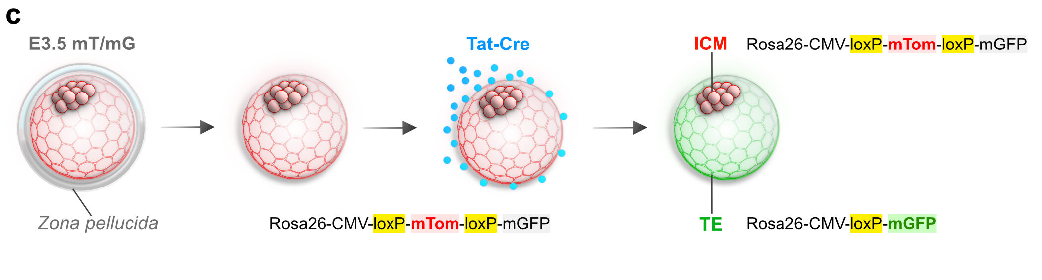
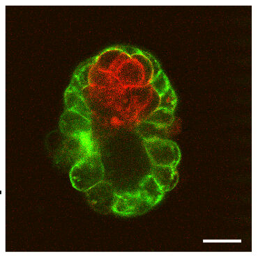
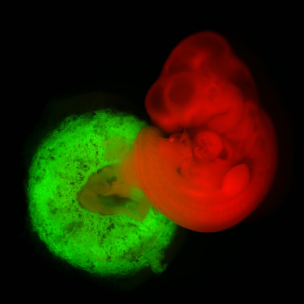
Tat-Cre mediated recombination (deletion) in the trophectoderm of mTom casette (red flourescence) and activation of mGFP expression
From a few hours of treatment to the placenta
The really useful feature of this approach is that the genetic manipulation happens at the blastocyst stage, when the trophoblast lineage is still being established.
Once recombination occurs in these early trophectoderm cells, the genetic change is inherited by their descendants.
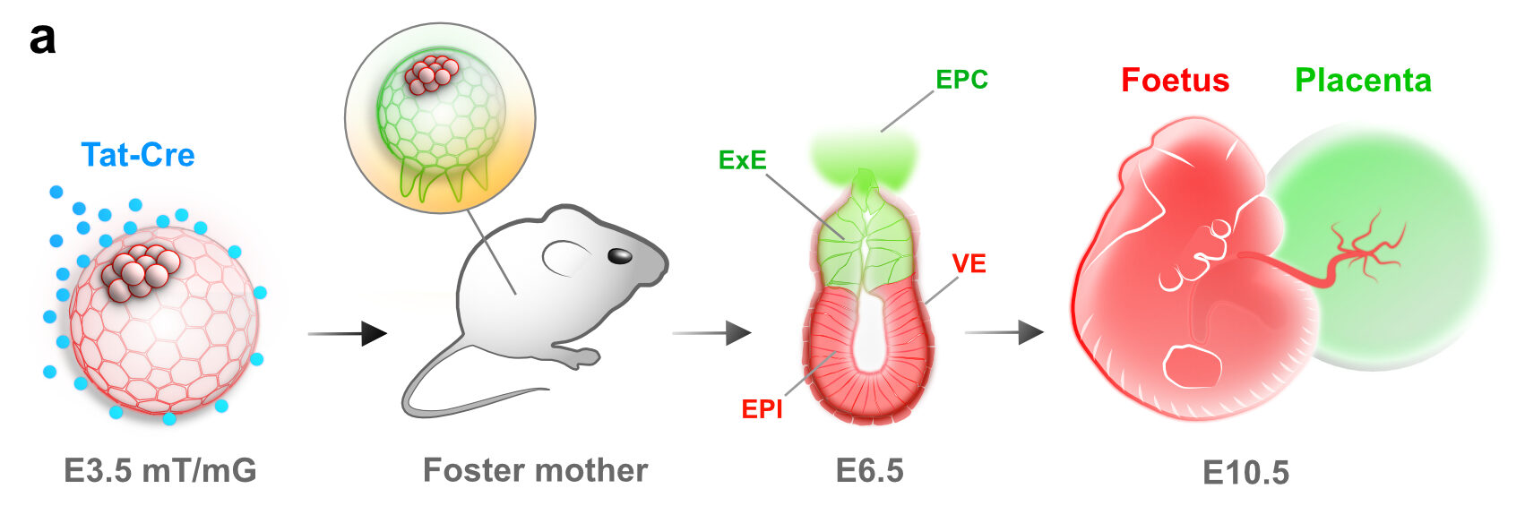
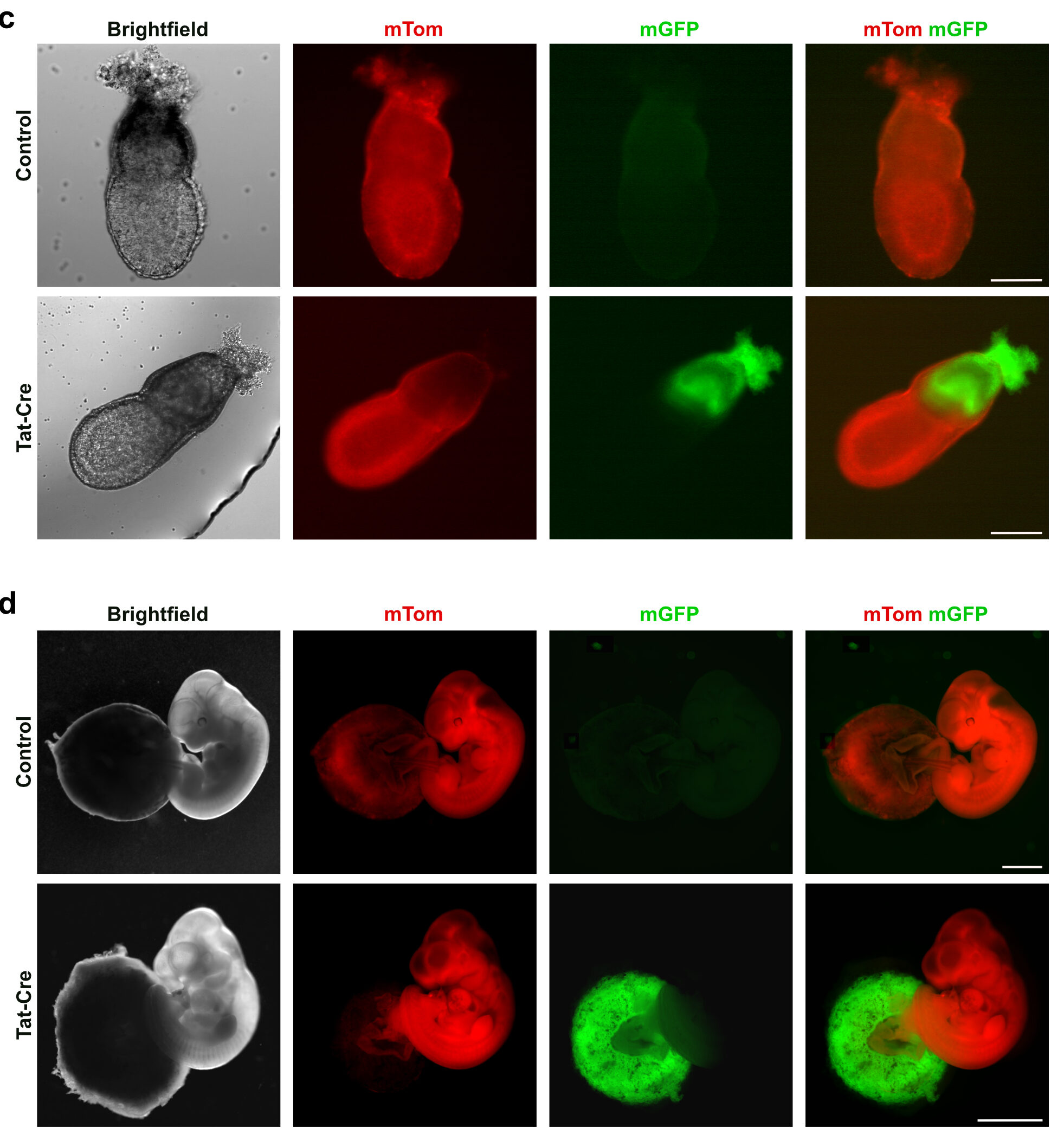
This means that a short treatment of an early embryo can produce a much longer-lasting effect: the altered cells go on to generate the trophoblast lineage during implantation and placental development.
We tested the approach using fluorescent reporters and conditional alleles and found that the method was efficient, rapid and did not compromise the developmental capacity of the treated embryos.
Why this matters for implantation
This is more than a technical shortcut.
Implantation is one of the most vulnerable stages of pregnancy. The trophoblast has to attach to the uterus, proliferate, differentiate and interact with maternal tissues to establish the placenta.
Defects in placental development are associated with embryonic abnormalities and pregnancy failure, yet it is often difficult to determine whether a developmental phenotype originates in the embryo itself or in the placenta.
A tool that allows us to manipulate the trophoblast independently gives us a way to ask much more precise questions:
Is this gene required by the embryo, the placenta, or both?
And perhaps even more importantly:
What happens to the embryo when the placenta is genetically altered?
This distinction is essential because the placenta does not simply support an already-developed embryo. It actively controls the environment in which the embryo grows.
A simpler route to conditional knockouts
Our approach also has practical advantages.
Traditional trophoblast-specific genetic models require the generation and breeding of additional Cre mouse lines. Tat-Cre avoids this step. We can take embryos carrying the appropriate conditional allele and introduce Cre directly at the blastocyst stage.
This makes the method relatively fast and straightforward, while also reducing the breeding required to generate the desired experimental embryos. Using homozygous conditional animals, for example, allows essentially all treated embryos to undergo the desired recombination rather than relying on the correct combination of alleles through breeding.
Importantly, the approach also avoids introducing a Cre transgene that could later become active in other tissues.
Giving the placenta its own genetic toolbox
One of the broader lessons from this work is that physical properties of the embryo can themselves be turned into experimental tools.
Instead of searching for a perfect promoter to drive Cre specifically in the trophoblast, we used something the embryo already provides: its natural permeability barrier.
A transient exposure to a cell-permeable protein was enough to exploit this difference and selectively manipulate the trophoblast lineage.
This gives us a direct way to investigate gene function during implantation and placentation and, more broadly, to separate the contributions of embryonic and extraembryonic tissues to development.
The placenta is often described as a support system for the embryo. But to understand embryonic development properly, we need to study the placenta as a developmental tissue in its own right.
Sometimes, the best way to understand the embryo is to change the cells that surround it.
In short
We developed a simple method for trophoblast-specific gene editing in the mouse blastocyst. By combining the natural permeability properties of the trophectoderm with cell-permeable Tat-Cre, we can activate Cre/loxP recombination in the trophoblast lineage without requiring a trophoblast-specific Cre mouse line.
This provides a rapid and flexible way to investigate gene function during implantation and placental development, and to disentangle the contributions of the placenta and embryo to normal and abnormal development.
A few hours of gene editing at the blastocyst stage can give us a window into the genetic mechanisms that build the placenta.
Read the research article:
Placental gene editing via trophectoderm-specific Tat-Cre/loxP recombination
Ozguldez HO, Fan R, Bedzhov I.
Development2020
https://doi.org/10.1242/dev.190371

You must be logged in to post a comment.