
How does a simple ball of cells transform into an organised embryo?
Around the time of implantation, the mouse embryo undergoes one of the most dramatic transformations in early development. The blastocyst, which contains a hollow cavity surrounded by several cell populations, begins to reorganise into an elongated structure called the egg cylinder. At the centre of this structure, the epiblast — the pluripotent tissue that will give rise to the entire embryo — transforms from a simple ball of cells into a cup-shaped epithelium surrounding a new fluid-filled space: the proamniotic cavity.
But where does this cavity come from?
And perhaps more importantly, what happens to the fluid that was already inside the blastocyst?
We set out to follow this process and discovered that the formation of the proamniotic cavity is a remarkably dynamic process involving cell polarity, cell–cell adhesion, fluid transport and cell division. Even more surprisingly, the cavity is not simply a by-product of tissue organisation. It becomes an important part of the mechanism that controls embryonic growth and patterning.
From one cavity to another
At the blastocyst stage, the embryo already contains a large fluid-filled space: the blastocoel.
The epiblast sits inside this structure as part of the inner cell mass. Following implantation, the epiblast begins to proliferate and expand. Rather than remaining inside the blastocyst, it progressively grows into the existing blastocoel and starts to reorganise into the egg cylinder.
This creates an intriguing situation.
The embryo already has a cavity, but it is about to create another one.
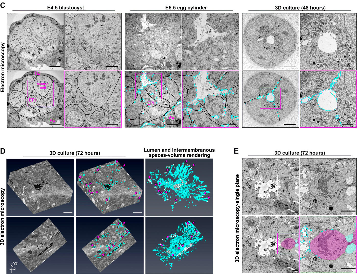
We found that these two cavities are physically connected through the process of development. As the epiblast expands into the blastocoel, fluid from the blastocoel flows into the newly emerging proamniotic cavity.
We could visualise this directly by injecting a fluorescent tracer into the blastocoel. At the blastocyst stage, the tracer remained within the blastocoel. But at later stages, as the egg cylinder formed, the same tracer appeared inside the proamniotic cavity.
This showed that the fluid filling the new cavity is supplied, at least in substantial part, by the pre-existing blastocoel. The embryo is therefore not simply making a new space independently. Instead, as its architecture changes, it effectively redirects an existing fluid compartment into a new developmental space.

How do cells make room for the cavity?
Knowing where the fluid comes from was only part of the story.
The next question was: how does fluid actually get between tightly packed epiblast cells?
At E4.5, the epiblast is a relatively compact ball of apolar cells. The cells are strongly connected to one another through E-cadherin-mediated adhesion.
As development progresses, this changes dramatically.
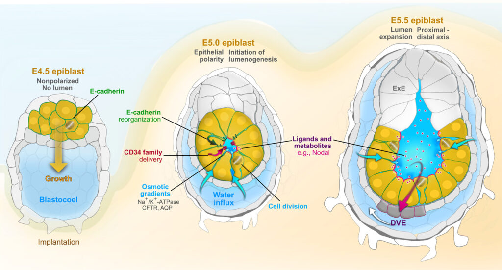
The epiblast begins to epithelialise and establish apical–basal polarity. The cells develop a new apical surface facing the future proamniotic cavity. At the same time, E-cadherin is removed from this newly specified apical domain, while members of the CD34 family of anti-adhesive proteins become enriched there.
The result is a local reduction in cell–cell adhesion precisely where the cavity will form.
This is important because fluid can now begin to separate the apical surfaces of neighbouring cells.
We think of this as a kind of hydraulic opening of the epiblast: fluid pressure acts on a region where cell adhesion has already been reduced, creating small intercellular spaces that can expand and merge into a continuous lumen.
The embryo uses pumps to move water
The fluid does not simply move passively.
Our experiments indicated that ion transport contributes to generating the pressure required for lumen formation. The coordinated activity of Na⁺/K⁺ and Cl⁻ transport systems, together with aquaporin-mediated water movement, drives vectorial fluid transport across the epiblast.
In other words, the epiblast behaves somewhat like a very small epithelial fluid-transport system.
Ions are moved across the cells, water follows, and the resulting pressure helps open the low-adhesion region at the centre of the tissue.
This provides a beautiful example of how cellular physiology can directly shape tissue architecture.
Cell division adds another push
There is another mechanism that makes the process even more interesting.
The epiblast is proliferating rapidly at this stage, and we found that mitosis itself contributes to lumen expansion.
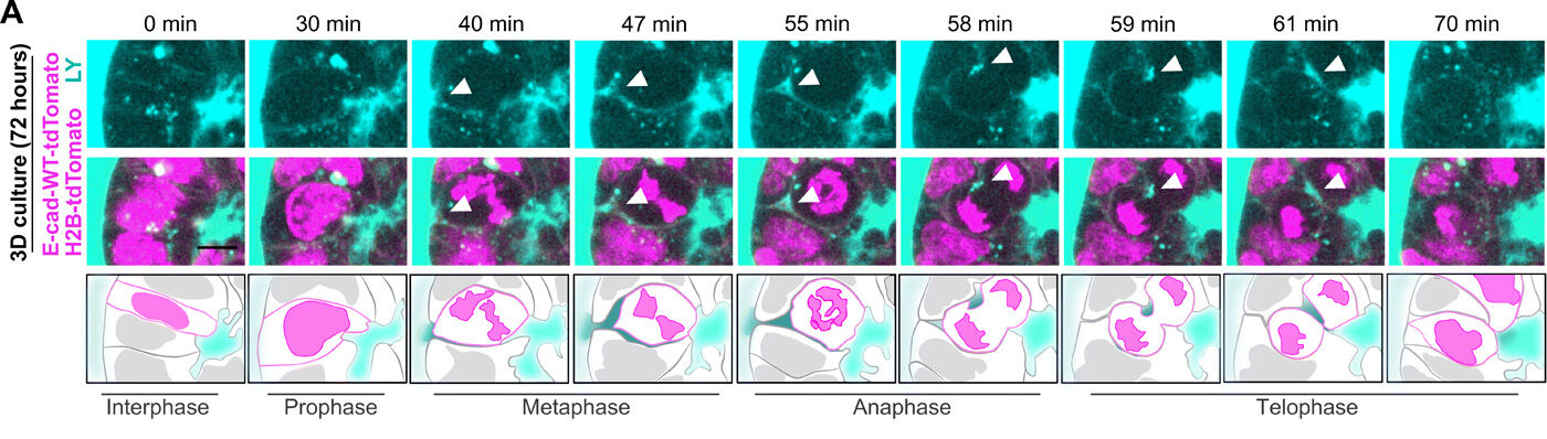
When an epiblast cell enters mitosis, its shape changes and the relationships between neighbouring cells are temporarily reorganised. During cell division, fluid can move through the spaces between neighbouring cells and around the dividing cell. We could follow this flow in live embryos and in our 3D stem-cell model.
As cytokinesis proceeds, fluid can also enter the space associated with the cleavage furrow.
These small pockets of fluid subsequently contribute to the growing lumen.
So cell division has a dual role: it increases the number of cells building the epiblast while simultaneously helping to expand the space at its centre.
A dynamic flow from the outside in
Putting these observations together gives us a very different picture of cavity formation.
The blastocoel surrounds the growing epiblast. As the epiblast epithelialises, its apical surfaces become less adhesive and begin to face one another. Ion transport generates fluid movement, while the physical rearrangements associated with cell division create additional routes for fluid to enter.
Blastocoel fluid therefore moves through the developing tissue and accumulates in the centre of the epiblast, where it becomes the proamniotic cavity.
The process can be thought of as:
blastocoel → intercellular spaces → emerging epiblast lumen → proamniotic cavity
This is not simply a passive leak of fluid. It is a coordinated process in which tissue polarity, adhesion, ion transport, water movement and proliferation work together to reshape the embryo.
The cavity starts talking
But why does the embryo go to all this trouble to make a cavity?
We found that the answer is not simply mechanical.
The proamniotic cavity becomes an important interface between the embryonic and extraembryonic tissues. When we experimentally interfered with lumen formation, the consequences extended beyond the cavity itself: embryonic growth and patterning were disrupted.
This suggested that the cavity acts as a developmental communication hub.
The surrounding tissues produce signals that influence the epiblast, including signals involved in establishing the anterior–posterior axis. By creating a central physical compartment, the cavity helps organise the spatial relationship between these signalling environments.
In this sense, the cavity does not just provide space for the embryo to grow.
It helps organise the conversations that tell the embryo how to grow.
Building the embryo by building space
This work changed the way we think about embryonic lumen formation.
It is tempting to view cavities as empty spaces that appear when cells organise themselves into tissues. Our findings suggest something much more dynamic.
The embryo starts with one cavity — the blastocoel. As the epiblast changes its polarity and architecture, fluid is redirected from this existing compartment into a new one. At the same time, adhesion is remodelled, ion transport generates fluid pressure and cell division helps drive fluid into the emerging lumen.
The result is the proamniotic cavity — a new space that then becomes an active participant in embryonic development.
The embryo builds the cavity, and the cavity helps build the embryo.
This provides a striking example of the self-organising nature of embryonic development: cells do not simply respond to pre-existing instructions. By changing their physical organisation and the spaces around them, they actively create new conditions that influence what happens next.
In short
We found that the proamniotic cavity forms through the coordinated action of epithelial polarisation, remodelling of cell–cell adhesion, vectorial ion and water transport, and cell division. Importantly, the fluid that fills the emerging cavity is supplied by the pre-existing blastocoel, creating a developmental flow from the original blastocyst cavity into the newly forming epiblast lumen.
Once formed, the proamniotic cavity becomes much more than an empty space. It provides a central environment for communication between embryonic and extraembryonic tissues and is required for proper embryonic growth and patterning.
The embryo does not simply make a cavity. It transforms one space into another — and in doing so, creates a new environment that helps determine its own development.
Read the research article
Deciphering epiblast lumenogenesis reveals proamniotic cavity control of embryo growth and patterning
Kim YS, Fan R, Kremer L, Kuempel-Rink N, Mildner K, Zeuschner D, Hekking L, Stehling M, Bedzhov I.
Science Advances 2022
https://doi.org/10.1126/sciadv.abe1640

You must be logged in to post a comment.