
Implantation is one of the most remarkable transitions in early pregnancy. The embryo has to attach to the uterus, invade the maternal tissue and establish connections with the mother’s blood supply. Yet many of the earliest events at this interface remain difficult to observe because the implanting embryo is buried deep within the uterine tissue.
We wanted to find a way to see what happens when the embryo first encounters the maternal vasculature.
Rebuilding the implantation environment
To do this, we developed a 3D biomimetic culture system that recreates key physical and cellular features of the mouse implantation site.
The system combines embryonic tissue with a three-dimensional extracellular matrix environment and maternal endothelial cells, allowing us to observe trophoblast invasion and embryo–blood vessel interactions directly.
This gave us something that is difficult to achieve in the uterus: the ability to watch the developing embryo and maternal vascular cells interact in real time and examine the molecular mechanisms underlying these interactions.
The embryo reaches out to the maternal vasculature
As implantation progresses, specialised trophoblast cells known as trophoblast giant cells leave the embryo and invade the surrounding tissue.
We found that these cells do not invade as isolated cells. Instead, they migrate collectively, forming penetrating strands that extend away from the embryo.
As they move through the surrounding tissue, the trophoblast cells acquire a surprising set of molecular features normally associated with blood vessels. They express vascular receptors, signalling molecules and adhesion proteins that allow them to communicate with maternal endothelial cells.
In this way, the invading trophoblast is not simply moving towards the maternal vasculature. It is actively acquiring the molecular machinery needed to find, recognise and communicate with blood vessels.
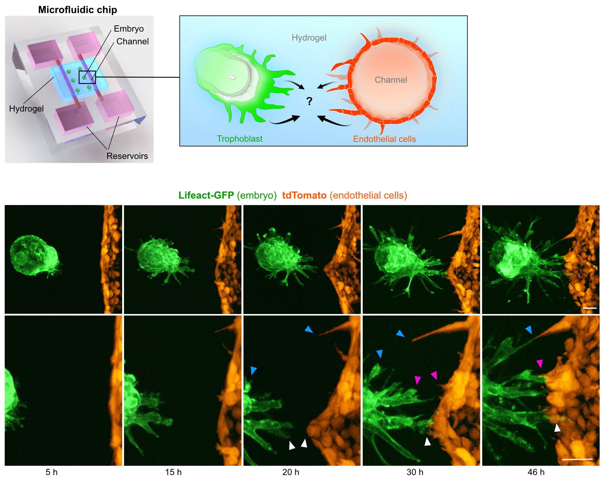
Mouse embryo and endothelial cells interactions in vitro, in a microfluidic chip
Building a communication network
We next asked what allows trophoblast cells and endothelial cells to establish contact.
By comparing the gene-expression programmes of trophoblast and endothelial cells, we identified a network of potential signalling interactions between the two cell types.
Among several angiogenic pathways that we investigated, PDGF signalling emerged as an important cue promoting the establishment of contacts between trophoblast and endothelial cells.
This suggests that the first interaction between the embryo and maternal vasculature is not simply a physical encounter. It is a highly coordinated process involving reciprocal molecular communication between the two tissues.
Seeing what is normally hidden
One of the advantages of our system is that it allows us to observe a process that is normally concealed by the uterus.
Inside the embryo, implantation happens in a complex three-dimensional environment containing many different cell types and extracellular matrix components. This makes it extremely difficult to isolate individual interactions and determine which signals control them.
Our biomimetic platform provides a simplified environment in which we can manipulate individual components and ask how they influence trophoblast invasion and vascular interactions.
It therefore provides a bridge between observing implantation in the embryo and testing its mechanisms experimentally.
Why does this matter?
The first connections between the embryo and maternal blood vessels are fundamental for establishing the future placenta and ensuring that the developing embryo can access the maternal circulation.
Our findings reveal that these interactions begin remarkably early. As trophoblast cells invade the maternal tissue, they collectively migrate and acquire a vascular communication programme that enables them to establish contacts with maternal endothelial cells.
More broadly, this work highlights how developmental processes can be understood by combining embryology with bioengineering. By recreating key features of the implantation environment, we can make hidden developmental events accessible to direct observation and experimental manipulation.
We developed a 3D biomimetic platform that allowed us to watch the first interactions between the embryo and maternal blood vessels and found that invading trophoblast cells actively build a molecular network for communicating with the maternal vasculature.
This provides a new way to study the earliest stages of embryo–maternal communication and the formation of the placenta.
Read the research article:
3D biomimetic platform reveals the first interactions of the embryo and the maternal blood vessels
Govindasamy N, Long H, Jeong HW, Raman R, Özcifci B, Probst S, Arnold SJ, Riehemann K, Ranga A, Adams RH, Trappmann B, Bedzhov I.
Developmental Cell 2021
https://doi.org/10.1016/j.devcel.2021.10.014

You must be logged in to post a comment.